At 3.0 mm pupil, the MTF values at 50lp/mm for the primary focus were 0.39 and 0.37, and for the secondary focus, 0.29 and 0.26 for the AT LARA and Proming IOLs, respectively. Light intensity assessment revealed that both IOLs allocate more energy to primary than secondary focus. The ray propagation of the two IOLs showed two distinct foci.
Itrace upgrade pro#
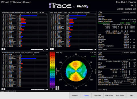
In addition, the optical quality of the IOLs was assessed by measuring the modulation transfer function (MTF) values at 50lp/mm and 3.0 and 4.5 mm apertures on the optical bench OptiSpheric® IOL PRO II. An experimental set-up with 0.01% fluorescein solution and monochromatic light (532 nm) was used to visualize the IOLs’ ray propagation. This study assessed the Proming EDOF Multifocal AM2UX and the AT LARA 829MP. To assess the optical behavior of a new diffractive intraocular lens (IOL) and compare its performance to that of an established extended-depth-of-focus (EDOF) IOL. Ray-tracing aberrometry can objectively assess accommodative amplitude in phakic eyes and pseudoaccommodation (depth of focus) in pseudophakic eyes. The young control group had the highest visual Strehl optical transfer function for near and distant targets (0.64 ± 0.24 and 0.56 ± 0.19, respectively), whereas the aberration-free IOL in the MX60E hyperprolate cornea group presented the lowest visual Strehl optical transfer function value for near (0.49 ± 0.19). There was no difference in effective range of focus, sphere shift accommodation, and pseudoaccommodations between the different IOL groups. The effective range of focus and sphere shift accommodation in the young control group were statistically larger than in the presbyopic group (P =. Sixty-two eyes received a Tecnis ZCB00, 39 a MX60E, and 43 a Tecnic ZXR00 Symfony IOL furthermore, 20 young phakic eyes and 19 presbyopic eyes were included in this study. Objective ray-tracing metrics of accommodation and pseudoaccommodation included the effective range of focus, sphere shift accommodation, and depth of focus. Ray-tracing wavefront analysis was performed 1 to 3 months postoperatively. The control groups consisted of young and presbyopic phakic patients. Patients with normal and hyperprolate corneas (post-hyperopic laser in situ keratomileusis) who underwent cataract surgery from March 2018 to October 2019 at the Medical University of South Carolina were examined and received either a diffractive intraocular lens (IOL) with an echelette design (Tecnis ZXR00 Symfony Johnson & Johnson Vision), a monofocal IOL with negative spherical aberration (Tecnis ZCB00 Johnson & Johnson Vision), or an aberration-free IOL (MX60E Bausch & Lomb). To compare objective measurements of accommodation and pseudoaccommodation in phakic and pseudo-phakic eyes using ray-tracing aberrometry. It is the investigation of choice in the today's era considering patient satisfaction and visual outcomes, postpremium intraocular lens implantations. The system integrates corneal topography with wavefront aberrometry, which has the unique feature of revealing the internal aberrations of the eye by subtracting the corneal aberrations from total aberration. This review will help all the ophthalmologists including residents and fellows learn the principle, features, and clinical applications of iTrace. In this review, our prime focuses on Ray Tracing aberrometer, iTrace. A variety of aberrometers with different principles are available, such as Ray Tracing, Hartmann-Shack, Tscherning, and automatic retinoscopy. Aberrometers are the most vital instruments used for estimating optical aberrations so that a more comprehensive understanding of optical error can be quantified and corrected. The QoV is primarily affected by both higher-and lower-order optical aberration.

Moreover, they are the departure of the performance of an optical system from the predictions of paraxial optics. Optical aberrations are defect in a lens or a mirror prevents light rays from being focused at a single point and results in a distorted or blurred image. Similarly, the quantity of vision is documented by measuring uncorrected and best-corrected distance visual acuities. The QoV is an integration of varied optical and neural factors. The quality of vision (QoV) can be easily assessed by documenting objectively the higher-order aberrations or by using subjective questionnaires available. A combination of both determines the final visual acuity. Visual acuity is the sum of qualitative and quantitative factors.


 0 kommentar(er)
